Labeled Dental X Ray Machine Parts

The Dental X Ray Machine Components And Functions Ppt Video Online Download

Tube Head And X Ray Tube Components Diagram Quizlet

Parts Of The X Ray Machine Diagram Quizlet
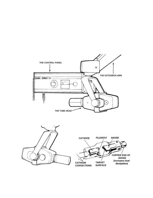
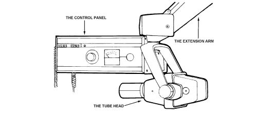
Dental X Ray Tube Head Diagram Find Local Dentist Near Your Area
Dental X Ray Equipment Parts Find Local Dentist Near Your Area
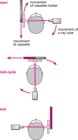
3 Panoramic Equipment And Imaging Pocket Dentistry

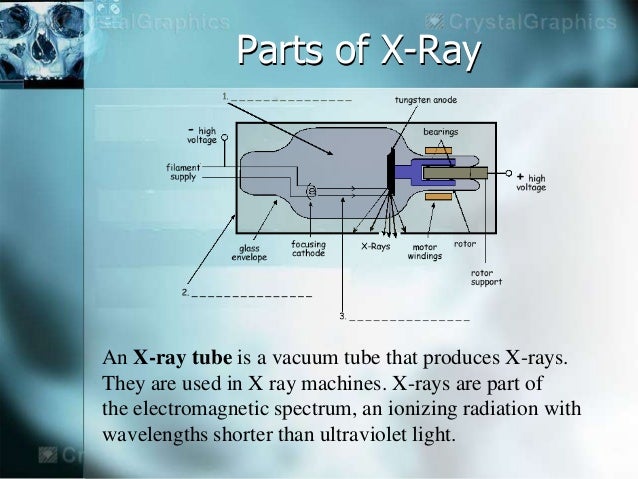
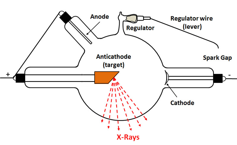
Anatomy of the x ray machine.
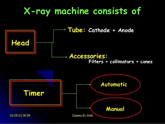
Labeled dental x ray machine parts.
Https Www Midmark Com Docs Librariesprovider2 Pdfs 00 02 1568 Rev V02 Jb 70 User Manual Pdf Sfvrsn 23bd0f47 4

Parts Of Dental X Ray Machine Flashcards Quizlet

Principles Of Dental Imaging Ppt Video Online Download

X Rays Definition Of X Rays By Medical Dictionary Radiology Humor Radiology Tech Radiology Student
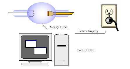
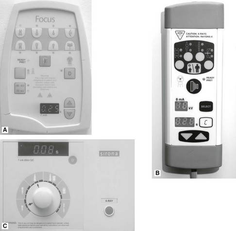
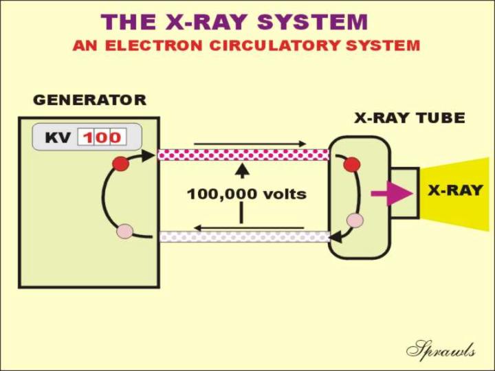
X Ray Generators
Dental X Ray Machine

Mw 8975 Chest X Ray Likewise X Ray Machine Diagram Likewise Schematic Diagram Download Diagram
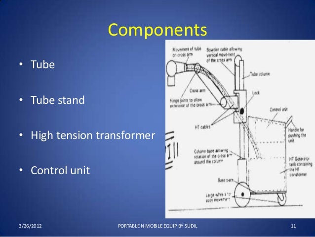
Portable N Mobile Unit
3 Dental X Ray Equipment Image Receptors And Image Processing Pocket Dentistry
Radiograph Sem
Https Www Midmark Com Docs Librariesprovider2 Pdfs 00 02 1608 Pdf Sfvrsn 47aec201 6

Eh 9435 Viewing Gallery For X Ray Machine Diagram Wiring Diagram

X Ray Production Animation Youtube

Cz 1251 Diagram X Ray Tube Collimators Integrated Circuit Diagram X Ray Wiring Diagram

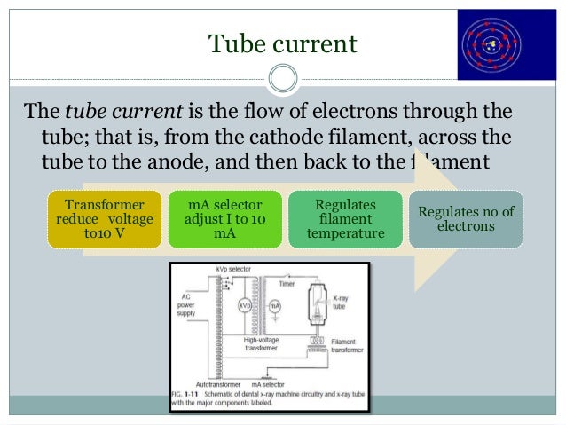
Chapter 6 Study Guide X Ray Tube X Ray Radiology Student
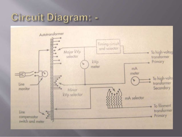
Introduction To The Parts Of X Ray Machine
1 Dental Radiology
Introduction To Gas Discharge Tubes And Cold Cathode X Ray Tubes
Https Encrypted Tbn0 Gstatic Com Images Q Tbn 3aand9gcrvlgd64hx9u5xj Alqjrdm7mcboh011n3hja6trypwrq9jza6s Usqp Cau
X Ray Production

Chapter 7 Dental X Ray Film Flashcards Quizlet

Coolidge X Ray Tubes General Information
X Ray Machine Ppt

Panoramic Radiograph Wikipedia
Source : pinterest.com
